


A genetic mutation
that causes albinism
in Doberman pinschers has been identified. And the researchers discovered that type of albinism has certain characteristics that are evident in humans too.
Paige Winkler, a doctoral student in the College of Veterinary Medicine at Michigan State University, and Joshua Bartoe, an assistant professor in the Department of Small Animal Clinical Sciences, led the effort to discover the mutated gene that is associated with a form of albinism in humans.
"What we found was a gene mutation that results in a missing protein necessary for cells to be pigmented," said Winkler. "Some defects in this same gene cause a condition called oculocutaneous albinism in humans. With an albino Doberman, you see a white or lighter-colored coat, pink noses and lips, along with pale irises in the eyes. These traits are very similar to the characteristics humans display with this particular condition causing light-pigmented skin and hair, along with eye discoloration and vision disturbances."
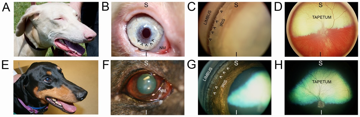
Partial Gene Deletion of SLC45A2 Causes Oculocutaneous Albinism in Doberman Pinscher Dogs. Images taken from WDP (top row) and black standard-color Doberman pinscher (bottom row). An image of WDP head (A) demonstrates lightly-pigmented nose, lips, and eyelid margins compared with the same darkly pigmented structures in SDP (E). A close-up image of WDP eye (B) shows: non-pigmented leading edge of the nictitating membrane (NM), tan-colored iris base transitioning to blue at pupillary margin, and oval-shaped dyscoric pupil aperture. The black arrowheads (in B) demarcate a region of significant iridal stromal thinning that was noted on examination to transilluminate (not shown in image) with retroillumination by light reflected from the tapetum lucidum. SDP eye (F) shows: darkly pigmented margin of the nictitating membrane (NM) and brown iris with a round pupil aperture. WDP gonioscopy image (C), which allows visualization of structures lying within the iridocorneal angle (in images C&G, this region lies between the words “LIMBUS” and “IRIS”) shows fibers of the pectinate ligament (demarcated by black arrowheads) are of a similar tan-color to the iris base, whereas fibers of the pectinate ligament (demarcated by white arrowheads) are dark brown in SDP (G). WDP fundus image (D) shows yellow-colored tapetum lucidum (labeled “TAPETUM”) and significant hypopigmentation of the retinal pigment epithelium and choroid allowing visualization of the choroidal vasculature. SDP fundus image (H) shows green-colored tapetum lucidum (labeled “TAPETUM”) and heavy pigmentation of the non-tapetal fundus. For orientation purposes, images taken at higher magnification (B–D and F–H) have the superior (S) and inferior (I) globe positions labeled.
doi:10.1371/journal.pone.0092127
The canine breed and people also experience the same skin sensitivity to sunlight, which results in an increased risk of skin tumors.
"We knew that the albino Dobermans typically developed these types of tumors, much like humans, but we wondered what the actual increase in prevalence was between a 'white' dog and a regular-colored Doberman," said Bartoe. "What we found was a significant increase in risk for development of melanoma-like tumors in the albino dogs."
Bartoe and Winkler studied 20 dogs of each color and discovered that more than half of the albino dogs had at least one tumor while only one of the regular-colored dogs possessed a tumor.
The results of their research could be of great importance to Doberman breeders around the world. Currently, American Kennel Club registration restricts dogs with the condition.
"Because Dobermans can carry the defective gene, but show no signs of the disorder, this has posed serious problems among breeders," said Bartoe. "But now that we've identified the mutation, we can look at the genetic makeup of these dogs and determine if they might be carriers."


Comments