


Computed tomography scans are the standard of care for diagnosing heart and lung conditions but there is worry more CT scans could mean a higher lifetime risk of cancer from radiation exposure.
A study, of 2,085 patients at nine centers in the U.S. and Middle East, found that using newer generation, dual-source CT scanners significantly reduced radiation exposure for patients when compared with first generation, 64-slice, single-source scanners or first generation, dual-source CT scanners.
Patient radiation exposure was reduced by 61 percent with the newer scanners, with no significant difference in image quality for patients having CT scans for coronary artery disease, pulmonary embolism or aortic disease.
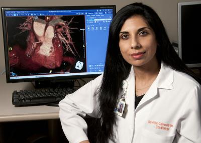
Kavitha Chinnaiyan, M.D., director of Advanced Cardiac Imaging Research at Beaumont Hospital, Royal Oak. Credit: Beaumont Health System
"Newer technology makes a difference in terms of radiation exposure and the difference is quite large," says study author Kavitha Chinnaiyan, M.D., director of Advanced Cardiac Imaging Research at Beaumont Hospital, Royal Oak. "It is important for patients to ask questions when referred for a radiation-based test to understand what the procedure involves and what the risks are of the particular technique and if there are alternative imaging choices."
The study findings also have important implications for referring physicians.
"Clinicians must understand that imaging studies not only have a major impact on the care of an individual patient, but also on trends in radiation exposure, as well as overall health care costs," says Dr. Chinnaiyan. "Incidental findings may require further imaging studies with other radiation-based tests. In addition to choosing patients appropriately, it is important to discuss the risks and benefits of testing with patients, and to refer them to centers that offer newer technologies."
The study results provide information that will help in setting standards for radiation safety quality control in cardiovascular imaging.
Beaumont cardiologists are world leaders in cardiac CT imaging research and have published studies on the use of CT imaging for identifying heart obstructions requiring invasive heart catheterization; for diagnosing or predicting heart attack; for identifying unstable plaque likely to cause a heart attack; and for diagnosing chest pain patients in the Emergency Center.


Comments