


A team of researchers from the Center for Imaging Science at the Johns Hopkins University and the CMLA of the École Normale Supérieure Cachan have demonstrated new algorithmic technologies for the parametric representation of human shape and form. Coupled with advanced imaging technologies, this presents opportunities for tracking soft-tissue deformations associated with cardiovascular studies, radiation treatment planning in Oncology, and neurodegenerative brain illnesses. The software algorithms provide tools for basic science and pre-clinical investigations for synchronizing structural and functional information across anatomical scales, allowing for the building of BrainClouds of physiological information in human brain mapping and thus connecting information across multiple anatomical and physiological scales.
"This is a remarkable combination of algorithm development and software technology that provides the basis for extending classical notions used for the positioning of rigid bodies adapted for the positioning of deformable bodies appropriate to human coordinates systems. Connecting this to modern machine learning algorithms opens the door for future research and clinical applications in which high throughput massive databases of structural and functional anatomy can be indexed and searched, analogous to the current state of the art in GoogleMaps", says Michael I. Miller Ph.D., a professor of Biomedical Engineering of the Johns Hopkins University and senior author on this paper.
By linking notions from Lagrangian and Hamiltonian mechanics of rigid bodies, the investigators have defined human shape as a Riemannian metric space generalizing D'Arcy Thompson's classical formulation of mathematical morphology of shape and form, with the metric structure defined by the geodesic flow of coordinates connecting one shape to another.
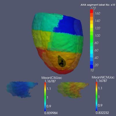
This is the Geodesic Positioning of 25 subjects in AHA atlas coordinates with colors representing AHA parcellation; black area located in anterior apical segment 13 showing structural phenotype difference between ischemic and non-ischemic cardiomyopathy at end-systole. Bottom shows smaller wall thickening during maximum contraction at end-systole at the location of infarction (segment 13) in ischemic population as signaled by Jacobian of geodesic coordinates indexed to the segment. Left: mean Jacobian for ischemic population, Right: mean Jacobian for the non-ischemic population. Note ischemic group has significantly smaller myocardial tissue volume. (Cardiac study done in collaboration with Dr. Robert G. Weiss, Director of DW Reynolds Clinical Cardiovascular Research Center in the division of Cardiology in the Johns Hopkins School of Medicine, and Dr. Siamak Ardekani. Data collected on an Aquilion 64(32) multi-detector computed tomography scanner, Toshiba Medical Systems Corporation, Japan; in plane resolution 0.36 × 0.36 mm (0.45 × 0.45 mm), out of plane thickness = 0.5 mm).
(Photo Credit: TECHNOLOGY journal)
"Once human shape is embedded in these Riemannian or geodesic coordinates, then human anatomy can be indexed and searched", Miller continued. "This is a beautiful example of how advances in imaging technology coupled to computational and algorithmic methods are enabling both biological discovery and clinical applications."
Using large volumes of spatial and temporal data being generated via high throughput imaging systems, the investigators in the United States and Europe are exploiting these geodesic positioning systems to uncover new biomarkers and diagnostic parameters that may provide clues to fundamental disease mechanisms in several neuropsychiatric illnesses, including dementia, Huntingdon's, schizophrenia, autism and mood disorder diseases.
At the same time another team from the Johns Hopkins School of Medicine, led by Drs. Susumu Mori and Thierry Huisman in collaboration with Jonathan Lewin, the director of the Department of Radiology, is deploying a Pediatric Brain Cloud.


Comments