


Researchers have found the earliest fossil evidence for the presence of bone marrow in the fin of a 370 million-year-old fish,
Eusthenopteron, a Devonian lobe-finned fish from Miguasha in Canada that is closely related to the first tetrapods.
Long bones, which are found in the limb of tetrapods, are not only important for locomotion and supporting the weight of the body, but also host the bone marrow. The latter plays a major role in hematopoiesis, i.e. the formation of blood cells. In a healthy adult human, about a hundred billion to one trillion new blood cells are produced every day to maintain the stable blood circulation.
The bone marrow also has an important role as part of the immune system. Although long bones are a rich source for marrow transplantation, the establishment the bone marrow in the medullary cavity and its interactions with the surrounding bone still remain partly mysterious and its evolution is not well understood.
Researchers from Uppsala University in Sweden and the European Synchrotron Radiation Facility (ESRF) in France decided to look for the origin of the bone marrow within vertebrates, using synchrotron microtomography to investigate the interior structure of fossil long bones without damaging them. They discovered that Eusthenopteron already exhibited typical marrow processes inside its humerus (upper arm bone).
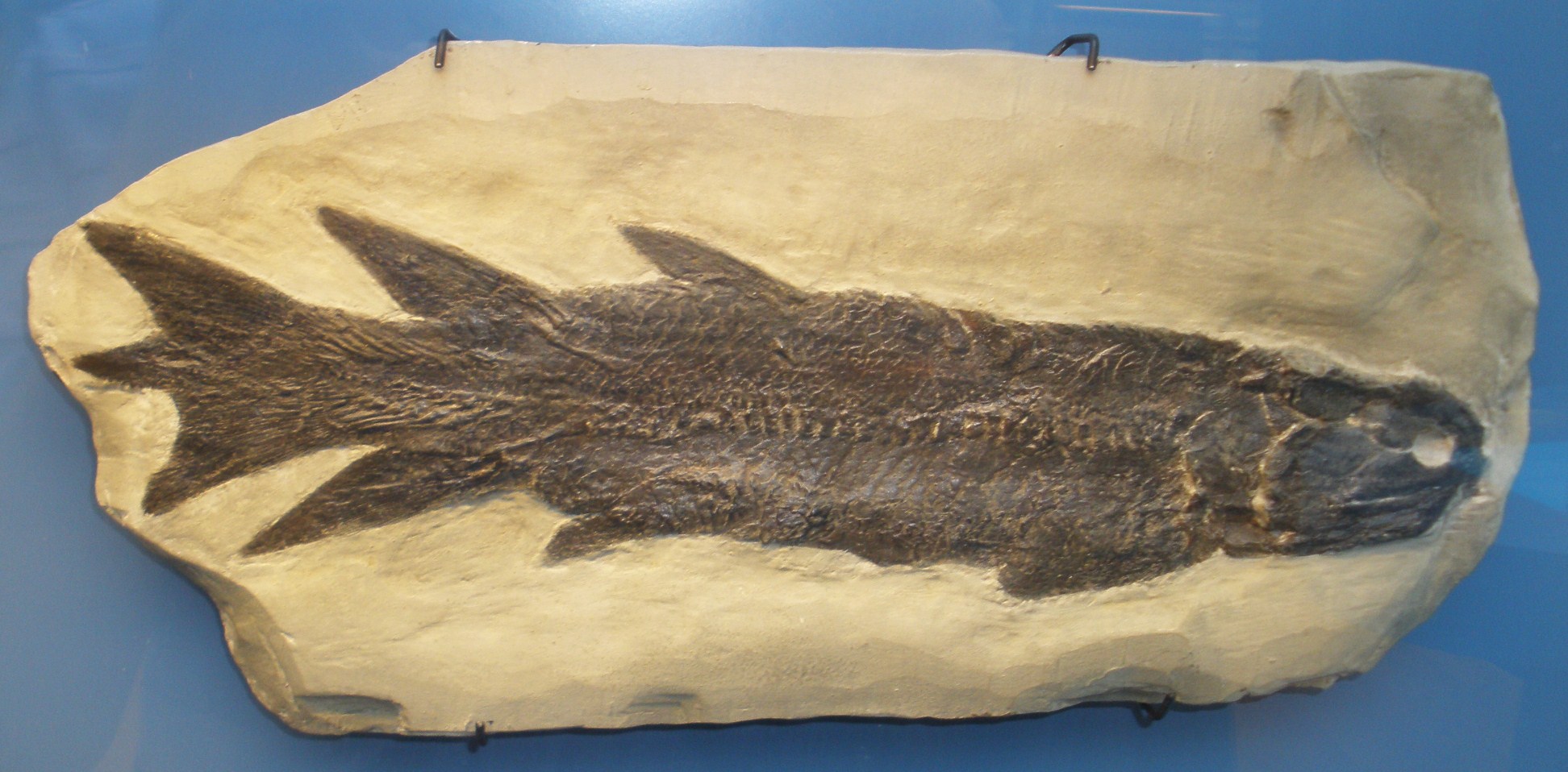
Eusthenopteron foordi. Credit: Wikipedia.
These processes are longitudinal, larger than blood vessel canals, and connect to the shoulder and elbow joint surfaces of the humerus. Thanks to the beam power of the ESRF, they were able to reach submicron resolutions and accurately reconstruct the 3D arrangement of the long-bone microanatomy of this close relative of tetrapods.
"We have discovered that the bone marrow certainly played a major role in the elongation of fin bone through complex interactions with the trabecular bone" says Sophie Sanchez, a researcher from Uppsala University and the ESRF. "This intimate relationship, which has been demonstrated by molecular experiments in extant mammals, is actually primitive for tetrapods".
This discovery is very important for understanding the evolutionary steps that built up the distinctive architecture of tetrapod limb bones and created a location for the distinctive, complex and functionally important tissue that is bone marrow. It is also a powerful demonstration of the capabilities of synchrotron microtomography.
"Without the 3D information provided by the synchrotron, we could never have understood the internal organization of the marrow space" says Per Ahlberg from Uppsala University. "If you cut a slice through a bone like this, which would damage it irreparably, you would only see an uninformative pattern of holes in the cut surface. With the synchrotron we can image the whole internal structure and understand how the marrow processes are organized, without doing any damage to the bone at all."
Source: Uppsala University


Comments