


Scientists at the U.S. Department of Energy's Brookhaven National Laboratory have developed a method for correlating the results of microscopic imaging techniques in a way that could lead to improved understanding, diagnosis, and possibly treatment of a variety of disease conditions, including Alzheimer's disease. The Laboratory has filed a U.S. provisional patent application for the invention.
The invention is essentially a micron-scale metallic marking grid upon which scientists place their samples - biological tissues or inorganic samples such as minerals - prior to imaging with different methods. "When the findings are analyzed, the grid can be used to 'map,' or orient, the images to one another, allowing us to study multiple variables in a single sample and better understand how they relate to one another," said biophysicist Lisa Miller, leader of the team that developed the new method.
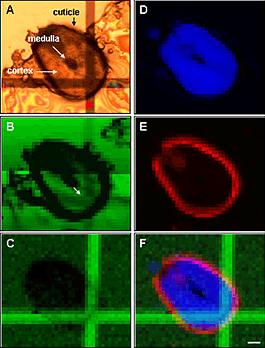
For example, many diseases such as Alzheimer's are characterized by changes in both organic materials, such as proteins, as well as changes in the composition or concentration of inorganic trace metals (e.g., iron, copper, and zinc). Scientists have techniques - infrared spectroscopy and x-ray fluorescence - for studying each of these independently. But without a way to correlate the findings from the two methods, important information about the relationship between the organic and inorganic components can be missed.
"The x-ray and infrared-sensitive grid allows for the study of both pathological symptoms by precisely overlapping the results of these imaging methods," Miller said. "This ability to correlate images will ultimately lead to a more complete picture of many disease states." The grid is deposited in two thicknesses onto an x-ray transparent material like mylar. It can be made of any metal, but gold is preferred. The "bars" of the grid are only a couple of nanometers thick, whereas the remainder of the metal surface is thicker. The dual thicknesses make the pattern sensitive to both infrared reflectivity and x-ray fluorescence imaging.
In another version of the invention, a single-layered grid is used to correlate light microscopy with x-ray fluorescence imaging.
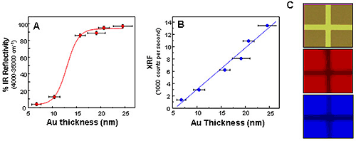
Once the images are collected, custom software uses the grid patterns to align the images and correlate them with each other.
In addition to helping scientists study disease processes, the method could also be applied in monitoring and/or cleaning up environmental contamination, which is also characterized by the interplay of organic and inorganic factors.
This work was performed at Brookhaven Lab's National Synchrotron Light Source (NSLS), a source of intense infrared, ultraviolet, and x-ray beams used for studying atomic level details of a wide range of materials from biological molecules to semiconductor devices. More information on this study can be found at: http://www.nsls.bnl.gov/newsroom/science/2007/09-Miller.htm .
This project was funded by the National Institutes of Health with operational support for the NSLS provided by the Office of Basic Energy Sciences within the U.S. Department of Energy's Office of Science.
- Brookhaven National Lab


Comments