


By identifying the molecular structure of a vital biological chemical, researchers may have solved a long-standing debate.
The controversy is about a form of enzyme called a heme (or haem, as in haemoglobin) at the center of which is an iron atom (Fe) called a 'ferryl' which becomes oxidized when a reacting heme is in an intermediate state called Compound I.
The question is whether this oxidation involves just an oxygen atom (O), or a hydroxyl group (OH). The difference being one hydrogen ion, or in other words, a proton.
Much has been written in the scientific literature about this ferryl heme, some scientists arguing that it carries a proton, while others have been equally adamant that no proton is present. Resolving this fundamental inconsistency has implications for understanding of oxidative processes within living cells, which is crucially important for drug development.
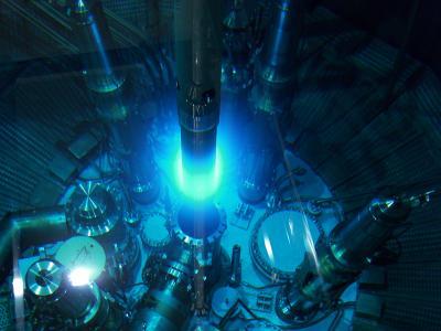
The core of the Munich reactor FRM II in use, showing the blue Cherenkov radiation. Copyright: J. Neuhaus / FRM II, TUM
Professors Peter Moody and Emma Raven from the University of Leicester, working with neutron crystallography experts from the Institut Laue-Langevin in Grenoble, the Maier-Leibnitz Zentrum (MLZ) near Munich, and the University of Manchester say they have solved the mystery of the ferryl heme Compound I structure using neutron crystallography.
"Finding out where all the hydrogens are is key to understanding the way enzymes work," said Professor Moody. "The ability to capture intermediates at cryogenic temperatures combined with the information available from neutron crystallography, means that we can finally see them."
Structures of proteins are commonly determined using X-ray crystallography, in which X-rays are fired at a crystalline form of the protein and the resulting diffraction pattern can be used to calculate the molecular structure. While this process works fine in general, it cannot be used to identify the location of hydrogen atoms for various reasons. Neutron protein crystallography, a technique originally developed in the 1960s, uses neutrons instead of X-rays – which eliminates these problems and enables the detection of individual hydrogen atoms. The team used neutron cryo-crystallography, which has the additional complication of using very low temperatures to cryo-trap the enzyme intermediate.
And the answer turns out to be … the ferryl heme in Compound I is NOT protonated. But, unexpectedly, the results showed that one of the amino acid side chains (a histidine) on the molecule IS doubly protonated, which raises questions of its own in terms of mechanisms for oxygen activation in heme enzymes.
"The exact structure of ferryl heme has been a long and difficult problem for the heme community to resolve," said Professor Raven. "Through our collaborators in Grenoble and Munich, we have been lucky to have had access to excellent neutron beamlines in Europe, which has meant that we were in an excellent position to try new and different approaches that worked very nicely. The single crystal EPR experiments at Manchester were essential to help us verify the stability of the ferryl species."
Article: 'Neutron cryo-crystallography captures the protonation state of ferryl heme in a peroxidase', Science.


Comments