Beliefs that tumors are caused by the modern world's food and chemicals have suffered a setback. A 60-million-year setback.
The fossilized tail of a young dinosaur from in southern Alberta, Canada had the benign tumor as part of the pathology of LCH (Langerhans cell histiocytosis), a rare and sometimes painful disease that still afflicts humans, particularly kids.
The first clue was the shape of cavities in the vertebrae of a tail of a young dinosaur of the grass-eating herbivore species. The large cavities in two of the vertebrae segments were extremely similar to the cavities produced by tumors associated with the rare disease LCH that still exists today in humans. Most LCH-related tumors suddenly appear in the bones of children aged 2-10 years. Thankfully, they disappear without intervention in many cases.
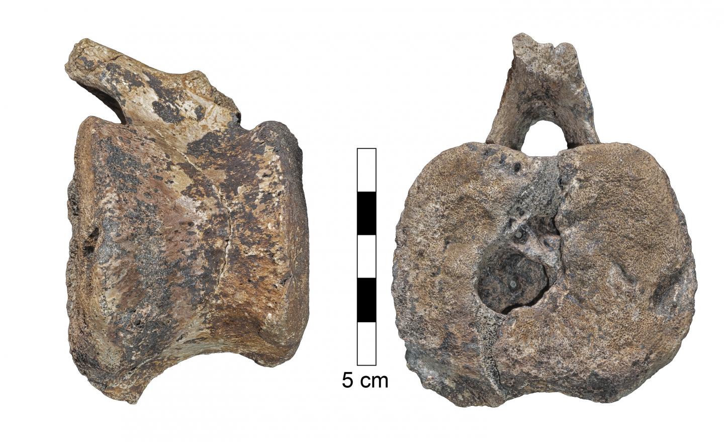
The larger hadrosaur vertebra in lateral view (left) and caudal view (right). The space that contained the overgrowth opens to the caudal surface of the vertebra. Credit: Assaf Ehrenreich, Sackler Faculty of Medicine, Tel Aviv University
The dinosaur tail vertebrae were sent for on-site advanced micro-CT scanning to the Shmunis Family Anthropology Institute at TAU's Dan David Center for Human Evolution and Biohistory Research, Sackler Faculty of Medicine, which is located at the Steinhardt Museum of Natural History.
They scanned the dinosaur vertebrae and created a computerized 3D reconstruction of the tumor and the blood vessels that fed it. The micro and macro analyses confirmed that it was, in fact, LCH. This is the first time this disease has been identified in a dinosaur.