


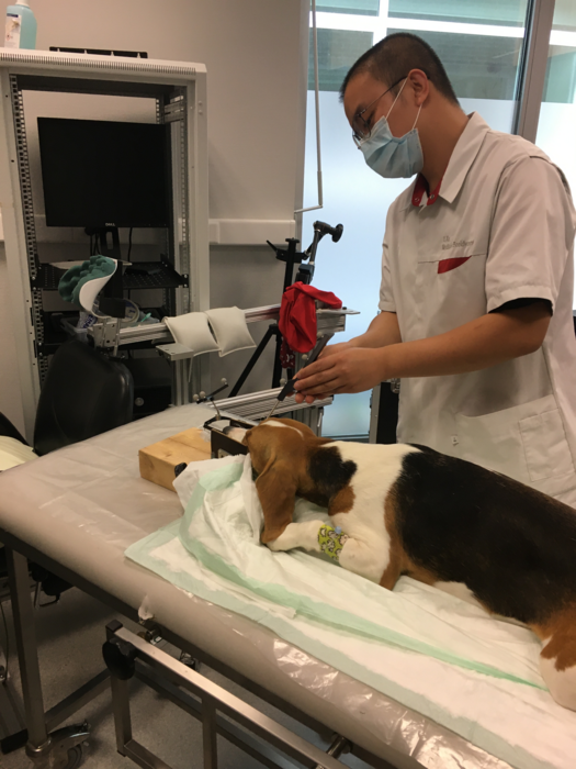
Animal models of anxiety can benefit both veterinary and eventually point to human medicine. However, the many different aspects of anxiety cannot all be studied effectively in the same animal model. Rodents are cheap and therefore often studied, but this new study takes advantage of the larger brains and bigger cortex found in dogs to characterize neural networks associated with anxiety. For the experiment, 25 healthy and 13 anxious good boys and girls were volunteered by their owners and examined via functional MRI (fMRI).
Credit: Xu et al., CC-BY 4.0 (https://creativecommons.org/licenses/by/4.0/)
Obviously the dogs were not harmed in any way, fMRI is just pretty pictures and scholars interpret how blood flow changes in them. The researchers compared the resting-state of dogs with and without anxiety, comparing network metrics and connectivity between groups, and determining their associations with anxiety symptoms. Resting-state fMRI indicated that functional connections between the amygdala and other parts of the anxiety circuit, particularly the hippocampus, were stronger than normal in anxious dogs. Within the anxiety circuit, network metrics including global and local efficiency were higher in the amygdala of anxious dogs. Dogs which exhibited fear and anxiety towards strangers, as well as excitability, were more likely to have brains showing abnormal network metrics in the amygdala.
The researchers believe their findings show that resting-state fMRI is a good tool for studying dog-models of anxiety, and that future studies like this could increase our understanding of how anxiety-related circuitry in the brain is altered in anxiety-disordered animals, and possibly even humans with the condition.


Comments