


In 1855, a specimen of the brain of mathematician Carl Friedrich Gauss was taken and preserved. But the over 150-year-old slice of his brain, which scientists had long been examining in the belief that it was Gauss's brain, turns out to not be his brain at all.
Instead, the preserved specimens of the brains of Gauss and Göttingen physician Conrad Heinrich Fuchs, a medical scholar and founder of the University of Göttingen's anatomical pathology collection, were switched, probably soon after the death of both men in 1855, says Renate Schweizer, a neuroscientist at Biomedizinische NMR Forschungs GmbH at the Max Planck Institute for Biophysical Chemistry.
She has now correctly identified the two brains, both of which are archived in a collection at the University Medical Center Göttingen, by use of a magnetic resonance imaging scanner.

The brains of Carl Friedrich Gauss and Conrad Heinrich Fuchs side by side. Wagner's lithograph of Fuchs's brain dating from 1862 (left) and his copperplate of Gauss's brain dating from 1860 (right) exhibit clear differences. The middle image is a recent MRI surface reconstruction of Gauss's brain. The divided central fissure of the left hemisphere is highlighted in yellow. Credit: Jens Frahm and Sabine Hofer / Biomedizinische NMR Forschungs GmbH 2013
Schweizer, a biologist and psychologist, made the discovery while working in her research field – the region of the brain around the so-called central fissure. The gyri running along the central fissure are where the brain processes stimuli, like touch, heat or pain, and where it controls movements. Renate Schweizer suspected that Gauss's brain featured a rare anatomical variation: a visible division of the central fissure. This is found in less than one percent of the population. Normally, it is of no significance to the people affected, though in a few cases it can cause minimal changes in motor and sensory function.
Schweizer spotted one of those central fissure divisions in the MRI scans believed to be of Gauss's brain, taken in 1998 by Jens Frahm and his team at Biomedizinische NMR Forschungs GmbH and searched through the primary literature to confirm her findings. Rudolf Wagner, an anatomist in Göttingen and friend of Gauss, had prepared the brain slices of both Gauss and Fuchs before studying them and documenting the images in publications dating back to 1860 and 1862. But contrary to what she expected to see, Schweizer did not find the divided central fissure in the images of Gauss's brain. Instead, her MRI images were a perfect match for Wagner's picture of Fuchs's brain.
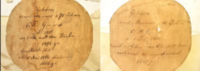
False labeling: Labels on the glass jars containing the brains of Gauss (left) and Fuchs (right). Credit: Böttcher-Gajewski / MPI for Biophysical Chemistry
When Schweizer visited the collection at the Institute of Ethics and History of Medicine, her initial suspicion was confirmed: the original brain taken from Gauss was indeed in a glass jar labelled 'C. H. Fuchs', while Fuchs's brain was in a jar marked 'C. F. G__ss'. "My theory, according to the information currently available, is that the brains were probably put into the wrong jars relatively soon after Wagner's examinations, at the time when the surface of the cerebral cortex was being measured again," says the neuroscientist.
After that, there were no further comparative studies of the brains of Gauss and Fuchs, which is why no one noticed the subsequent mix-up. It is also significant for the Göttingen-based Gauss Society to know that the brains of Gauss and Fuchs are now assigned to their proper owners once more. "The Gauss Society's Director, Axel Wittmann, was an active supporter of the project from the start and his extensive knowledge was extremely helpful in uncovering the mistake made so many years ago," reports Schweizer.
Her discovery shows how important historic collections are for modern-day research. Schweizer confirms: "It's a stroke of luck that the brains in the collection, which are in perfect condition, are still accessible to researchers more than 150 years down the line." That is what enabled the mix-up to be identified without a shadow of a doubt and the historical brains to be examined in the MRI scanner. Schweizer collaborated closely with former team colleague Gunther Helms, who works with brain slice MRIs in the MR Research Service Unit at the Department of Cognitive Neurology at University Medical Center Göttingen. As Jens Frahm, Director of Biomedizinische NMR Forschungs GmbH, emphasises: "We are not looking for the genie in the gyri of the brain. What we are most interested in is documenting specimens for the long term future to provide a foundation for continuing basic research." All MRI images and photographs of the historic brains are therefore being digitally archived, thus protecting them as long-term scientific assets. They are a significant impetus for new research projects: Schweizer herself is currently using the MRI images to study the divided central fissure in Fuchs's brain both above and below the surface of the cerebral cortex.
The MRI images also enable the scientists to demonstrate that earlier publications on what was believed to be Gauss's brain did not contain incorrect information. In those works, the mathematician's brain was described as normal. Walter Schulz-Schaeffer, who is head of the Prion and Dementia Research Unit of the Institute of Neuropathology at University Medical Center Göttingen, made a first examination of the recent MRI images and was able to confirm that the brain of the brilliant mathematician and astronomer Gauss, like that of the physician Fuchs, is largely anatomically unremarkable. The two organs are also similar in size and weight. "The age-related changes in Gauss's brain are normal for a man of 78. Changes in the basal ganglia are indicative of high blood pressure," comments the neuropathologist.
Not every MRI scan of a historical slice allows for such a clear assertion. That is why neuropathologists and MRI scientists are currently working together to study how tissue and organs change as a result of decades or centuries of storage in alcohol, and how adapted MRI methods can improve the interpretation of the images obtained.
The historical brains have, meanwhile, again found their well-earned rest in the university collection – with no chance of a mix-up ever again.


Comments