


3-D images are very useful in medicine and now they're gaining ground in physics. Researchers from Hahn-Meitner-Institute (HMI) and the University of Applied Science in Berlin have succeeded in creating a direct, three-dimensional visualization of magnetic fields inside solid, non-transparent materials for the first time.
This could prove invaluable because to understand high temperature superconductivity it is vital to understand how magnetic flux lines are distributed and how these flux lines can be established in materials. With this new experimental setup, it is now possible to visualize magnetic domains in magnetic crystals three-dimensionally.
The researchers in the imaging group used neutrons, subatomic particles that have zero net charge, but do have a magnetic moment, making them ideal for investigating magnetic phenomena in magnetic materials.
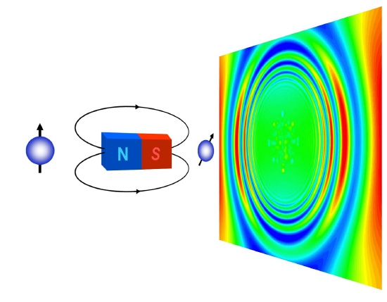
When in an external magnetic field, the neutrons behave like compass needles, all aligning to point on the direction of the field. Neutrons also have an internal angular momentum, often referred to by physicists as spin, a property that causes the needle to rotate around the magnetic field, similar to the way in which the Earth rotates on its axis.
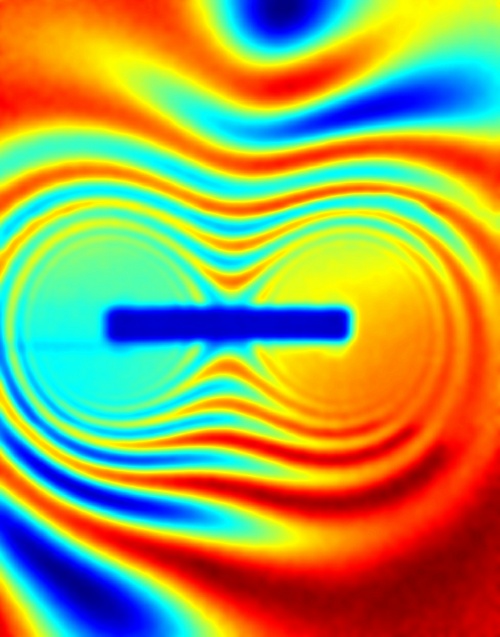
When all of the magnetic moments point in the same direction then the neutrons are said to be spin-polarised. If a magnetic sample is irradiated with such neutrons, the magnetic moments of the neutrons will begin to rotate around the magnetic fields they encounter in the sample and the direction of their spin changes.
Nikolay Kardjilov's group used this phenomenon as a measurement parameter for tomography experiments using two spin polarisers (which only allow the passage of neutrons whose spin points in a specific direction) to polarise and then analyse the neutrons. By detecting changes in the spins, it is possible to “see” the magnetic fields within the sample.
Kardjilov explains this by comparison with a medical CT scan; when a specimen is irradiated with x rays the density of the materials present alters the intensity of the light. "It's the same with our magnetic specimen, which changes the spin rotation of the neutrons", says Nikolay Kardjilov. "The equipment only allows passage of neutrons with a specific spin rotation, and this generates the contrast according to how the magnetic properties are distributed within the specimen. By rotating the specimen we can reconstruct a three-dimensional image."
Since 2005, Kardjilov has built up the neutron tomography section at HMI and now his group is the first to use spin rotation as a measurement signal for three-dimensional imaging. Normally, neutron imaging relies on the different levels of absorption of radiation by different materials to produce contrast.
The measurement of magnetic signals is a novel concept and its success lies partly in the polarisers and analysers, and the detector system, which have been developed and built by the HMI researchers.
Citation: Nikolay Kardjilov, Ingo Manke, Markus Strobl, Andre Hilger, Wolfgang Treimer, Michael Meissner, Thomas Krist and John Banhart, 'Three-dimensional imaging of magnetic fields with polarized neutrons', Nature Physics advance online publication: 30 March 2008


Comments