Stem cells, the body's wonder tool, are extremely versatile. They can develop in 220 different ways, transforming themselves into a correspondingly diverse range of specialized body cells.
Biologists and medical scientists plan to make use of this differentiation ability to selectively harvest cardiac, skin or nerve cells for the treatment of different diseases. However, the stem cell culture techniques practiced today are not very efficient. What proportion of a mass of stem cells is transformed into which body cells? And in what conditions?
“We need devices that keep doing the same thing and thus deliver statistically reliable data,” says Professor Günter Fuhr, director of the Fraunhofer Institute for Biomedical Engineering IBMT in St. Ingbert.

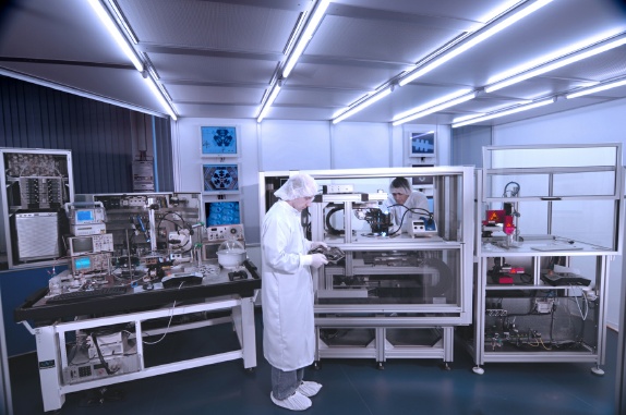
Two prototypes of laboratory devices, the result of the international project ‘CellPROM’ – ‘Cell Programming by Nanoscaled Devices’ – which was funded by the European Union and coordinated by the IBMT, may be the answer. Using these new tools, the development of stem cells can now be systematically observed and investigated with unprecedented accuracy.
“The type of cell culture used until now is too far removed from the natural situation,” says CellPROM project coordinator Daniel Schmitt – for in the body, the stem cells come into contact with solute nutrients, messenger RNAs and a large number of different cells. Millions of proteins rest in or on the cell membranes and excite the stem cells to transform themselves into specialized cells.
“We want to provide the stem cells in the laboratory with a surface that is as similar as possible to the cell membranes,” explains Daniel Schmitt. “To this end, the consortium developed a variety of methods by which different biomolecules can be efficiently applied to cell-compatible surfaces.”
The two machines – MagnaLab and NazcaLab – bring the the stem cells into contact with the signal factors in a pre-defined manner. In MagnaLab, several hundred cells grow on culture substrates that are coated with biomolecules. In NazcaLab, large numbers of individual cells, washed around by a nutrient solution, float along parallel channels where they encounter micro-particles that are charged with signal factors.
“We use a microscope and a camera to document in fast motion how individual cells divide and differentiate,” says Schmitt. The researchers demonstrated on about 20 different cell models that the multi-talents can be stimulated by surface signals to transform themselves into specialized cells.


Comments