


![]() We've known for some time that the islet is sensitive to a glucose demand in secreting insulin and uses gap junctions as a tuning parameter in this adaptation. Thus restoring natural cellular function through the use of microcurrents or electrical connectivity holds much promise for the future of diabetes treatment and pancreatic islet cell repair.
We've known for some time that the islet is sensitive to a glucose demand in secreting insulin and uses gap junctions as a tuning parameter in this adaptation. Thus restoring natural cellular function through the use of microcurrents or electrical connectivity holds much promise for the future of diabetes treatment and pancreatic islet cell repair.
Gap junction expression and coupling strength are very likely to occur as heterogeneous across the islet. If the naturally heterogeneous nature of gap junctions is acknowledged, this could be critical in designing appropriate clinical diabetes interventions.
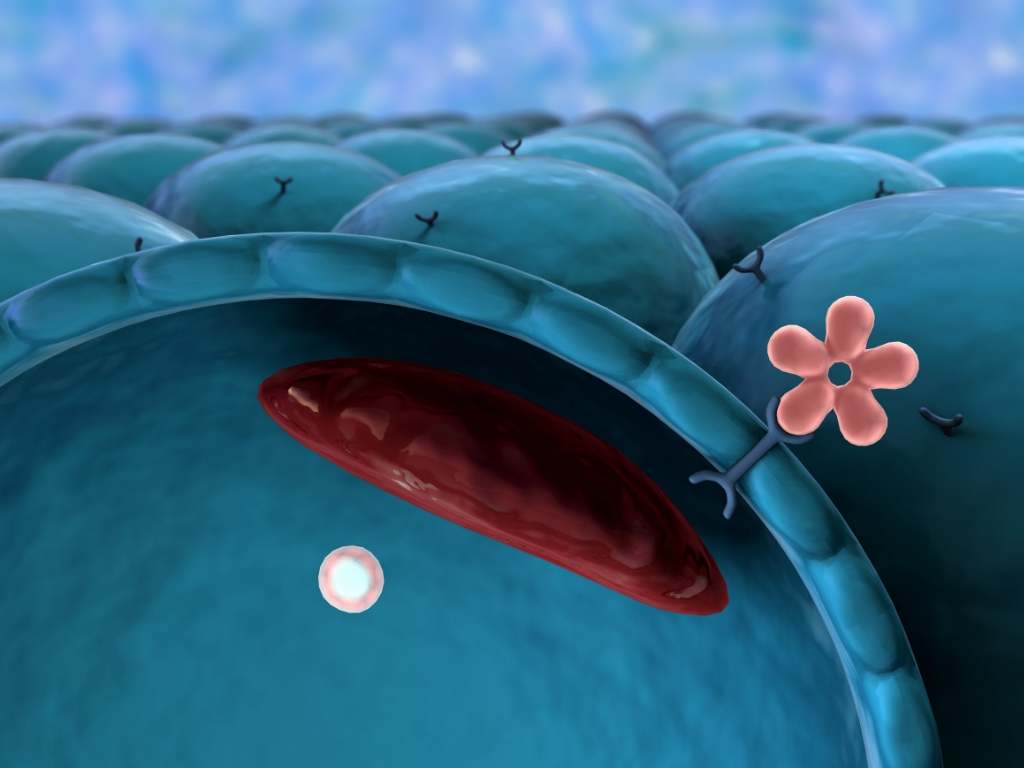
Gap junctions are a specialized intercellular connection between a multitude of animal cell-types. They directly connect the cytoplasm of two cells, which allows various molecules, ions and electrical impulses to directly pass through a regulated gate between those cells. Besides excitable cells, gap junctions are found between cells in almost every solid tissue [1]. From the cellular to the body systems, gap junctions are central to multicellular life [2], with numerous diseases linked to connexin disorders [3], including type 2 diabetes mellitus [4]–[6].
The islets of Langerhans in the pancreas are clusters of largely alpha-, beta- and delta-cells that respectively control secretion of the hormones: glucagon, insulin and somatostatin, central to energy regulation. Working in unison, they help maintain normal blood glucose (sugar) and salt levels in the body. Problems in the production or regulation of pancreatic hormones can cause complications related to blood sugar imbalance. Without this balance, we become susceptible to diabetes and related disorders. Gap junctions form direct connections between beta-cells [6], [10], [11] in islets, and are important for normal glucose-stimulated insulin secretion (GSIS) [7]–[9].
While we have expanded our understanding of the role and function of gap junctions and electrical conductivity in the healthy function of islet cells and established that microcurrents are a productive form of diabetes therapy, we had yet to see a treatment option come to market. It appears that this gap in treatment options has now been filled.
Cell MedX have produced the first device that mimics the endogenous electrical currents imperative to the healthy function of islet cells. The introduction of the eBalance device will bring to market the first non-invasive treatment option to capitalize on our understanding of how central microcurrents are to both treatment and potential reversal of type 2 diabetes. The eBalance produces a specific set of bioelectric signals that send pulses targeted at insulin producing beta cells and the protein, glucose transporter type 4 (GLUT4) contained therein. These pulses reset the natural gap junction, helping the cells to restore homeostasis.
Cells of almost all solid tissues are connected with gap junctions which permit the direct transfer of ions and small molecules, integral to regulating coordinated function in the tissue. The pancreatic islets of Langerhans are responsible for secreting the hormone insulin in response to glucose stimulation. Gap junctions are the only electrical contacts between the beta-cells in the tissue of these excitable islets. It is generally believed that they are responsible for synchrony of the membrane voltage oscillations among beta-cells, and thereby pulsatility of insulin secretion.
Most attempts to understand connectivity in islets are often interpreted, bottom-up, in terms of measurements of gap junctional conductance. This does not, however, explain systematic changes, such as a diminished junctional conductance in type 2 diabetes. This deficit is addressed via the model presented here, which is a learning theory of gap junctional adaptation derived with analogy to neural systems. Here, gap junctions are modelled as bonds in a beta-cell network, that are altered according to homeostatic rules of plasticity. A study by Pranay Goel and Anita Mehta, published in the peer reviewed journal PLOS, provides a compelling look at how learning theories reveal loss of pancreatic electrical connectivity in diabetes as an adaptive response. Gap junctions are clusters of intercellular channels between cells formed by the membrane proteins connexins (Cx), that mediate rapid intercellular communication via direct electric contact and diffusion of metabolites [1]. In excitable cells such as neurons, cardiac myocytes and smooth muscles, gap junctions provide efficient low-resistance pathways through which membrane voltage changes can be shared across the tissue.
Besides excitable cells, gap junctions are found between cells in almost every solid tissue [1]. Gap junctions are thus central to multicellular life [2], with numerous diseases linked to connexin disorders [3], including type 2 diabetes mellitus [4]–[6]. The islets of Langerhans in the pancreas are clusters of largely alpha-, beta- and delta-cells that respectively control secretion of the hormones glucagon, insulin and somatostatin central to energy regulation. Working in unison, they help maintain normal blood glucose (sugar) and salt levels in the body. Problems in the production or regulation of pancreatic hormones can cause complications related to blood sugar imbalance. Without this balance, we become susceptible to diabetes and related disorders. Gap junctions form direct connections between beta-cells [6], [10], [11] in islets, and are important for normal glucose-stimulated insulin secretion (GSIS) [7]–[9].
Gap junctions are generally believed to be important for coordinating the beta-cell electrical oscillations known as bursting, which in turn, can then support pulsatile insulin secretion [6], [10], [12]; this view is supported by previous studies [13]–[15].
The conductance strength of gap junctions evolves by the insertion or deletion of connexin proteins (Fig. 1) into junctional plaques, and by altering the single-channel conductance and probability of channel opening [1]. Whether these molecular changes constitute a systematic adaptive response of the endocrine tissue to its metabolic environment remains to be investigated, in particular from a theoretical point of view.

Figure 1. Gap junctions between cells permit intercellular communication. Gap junctions are a specialized intercellular connection between a multitude of animal cell-types. They directly connect the cytoplasm of two cells, which allows various molecules, ions and electrical impulses to directly pass through a regulated gate between cells. Figure credit: Mariana Ruiz LadyofHats, http://en.wikipedia.org/wiki/File:Gap_cell_junction_en.svg. https://doi.org/10.1371/journal.pone.0070366.g001
As with many other excitable cells, the information content of bioelectric signals [16] in islets is yet unclear. The mechanisms underlying bursting are well understood [17], [18]; however, how those temporal properties regulate energy homeostasis is not. While slow (5–15 minute period) bursts are generally thought to drive secretion at stimulatory concentrations of glucose, faster (periods less than 5 minutes) oscillations are also found, typically at sub-stimulatory (basal) glucose levels; the average calcium signal, however, is comparable in either case (such as in simulations from [17], not shown). The hypothesis that a synchronous bursting of beta-cells [10], [12], [19] is essential to GSIS is guided by the observation of pulsatile insulin secretion from islets [20] and in vivo [21]. Gap junctions can certainly mediate synchrony in principle, as shown in both simulations [12], [13] and experiments [5], [22]. Stozer et al. [25] have recently demonstrated that in islet slices only local synchronization is seen across groups of beta-cells. Another theory, different from one that anticipates gap junctions serve to synchronize an islet uniformly, thus appears to be necessary to explain some of the phenomena associated with insulin secretion.
A paradigm that is gaining increasing recognition is that bioelectric and (epi-) genetic signaling are related as a cyclical dynamical system [16]: membrane voltage activity induces changes in mRNA expression and transcriptional regulation, which in turn leads to altered membrane channel proteins.
Goel and Mehta looked at an adaptive response of gap junctions to islet firing activity. Bioelectric cues are encoded as bursting, these determine junctional conductance states, and junctions respond in turn by translation modifications that alter firing rates. In this way, electric and genetic components “learn” from each other, iteratively. While learning is integral to neural systems and functionally beneficial at the level of a single individual, many studies have focused on the collective effects of [simple forms of] individual learning and decision-making, e.g. in populations of interacting individuals, or agents. Such distributed systems, exemplifying social or ecological group behavior, also share similarities with interacting systems of statistical physics, in the nature of the local “rules” followed by the individual units as well as in the emergent behavior at the macro level. Game-theoretic approaches [26]–[28] are sometimes brought to bear on such issues, their underlying idea being that the behavior of an individual (its “strategy”) is to a large extent determined by what the other individuals are doing.
The strategic choices of an individual are thus guided by those of the others, through considerations of the relative “payoffs” (returns) obtainable in interactive games. In this context, a stochastic model of strategic decision-making was introduced in [29], which captures the essence of the above-stated notion, i.e. selection from among a set of competing strategies based on a comparison of the expected payoffs from them. Depending upon which of the available strategic alternatives (that are being wielded by the other agents) is found to have the most favorable “outcome” in the local vicinity, every individual appropriately revises its strategic choice.
Voltage Gating of Junctional Conductance and Homeostatic Adaptation
Gap junctions are known to adapt on at least two timescales: trans-junctional currents are gated on a fast timescale of the order of a few milliseconds to seconds in response to a transjunctional voltage difference () [1]. Voltage gated currents of Cx36 channels (the connexin isoform relevant to islets [5]), expressed in Xenopus oocytes and transfected human HeLa cells, were recorded in [32] (Fig. 2). Haefliger et al. [33] have shown hyperglycemia decreases Cx expression in adult rats. Paulauskas et al. [34] have recently described a 16-state stochastic model of gap junctional currents that are voltage gated by altering, amongst other things, unitary single channel conductance and the probability of opening [1], [35]. On much slower timescales of hours to days, gap junctions are regulated by the events that alter the insertion and deletion of channels in the junctional plaque, connexin proteins synthesis, trafficking to the membrane and degradation. We propose to study adaptation in gap junction strength on slow timescales; this is the natural setting for a mean field theory of gap junction modification, that is, over suitably long periods that averages over cellular firing rates can be treated as adiabatic.
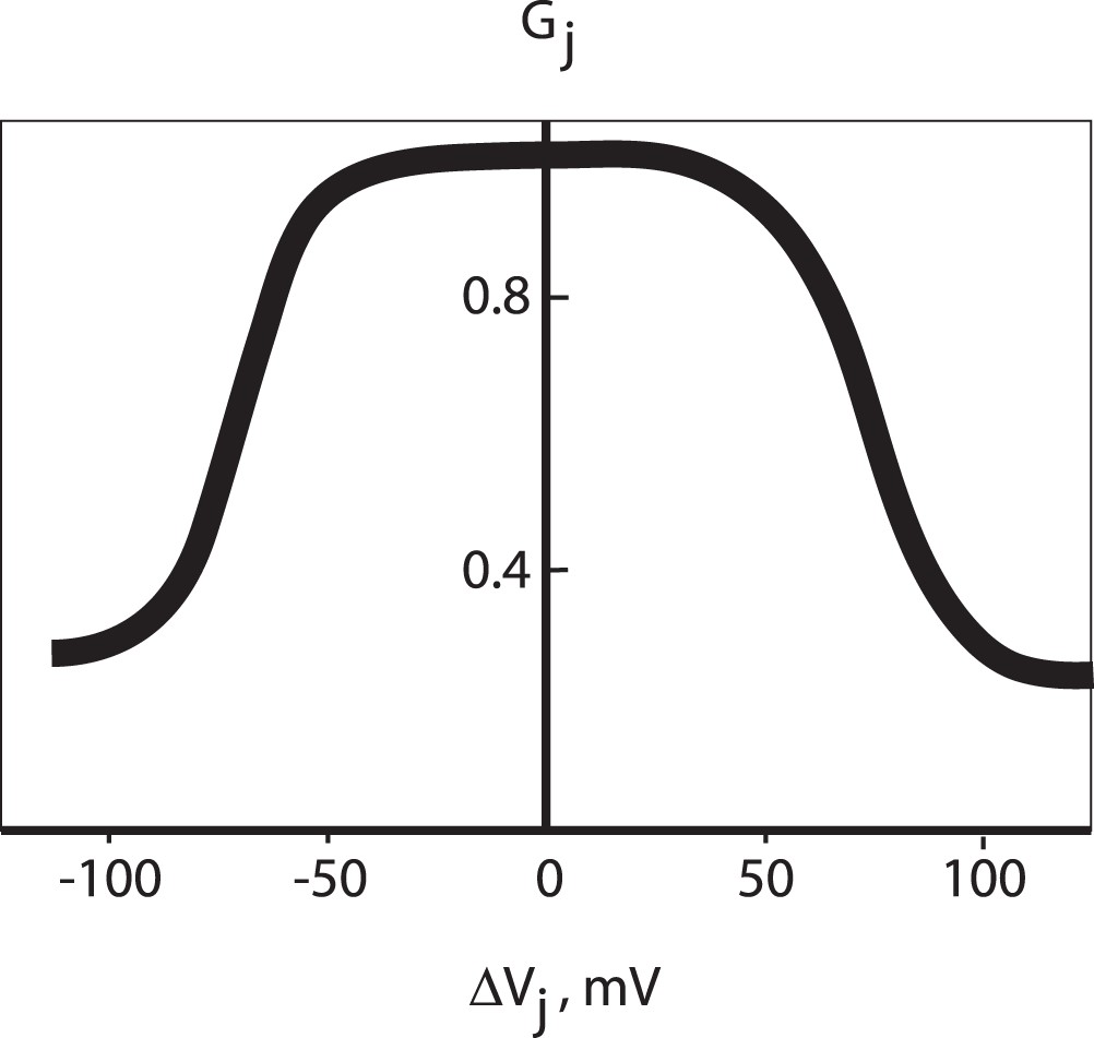
Figure 2. Voltage gating of Cx36 gap junctions, adapted from [32].
Steady-state junctional currents from HeLa-Cx36 cell pairs indicate conductance, , varies with transjunctional potential difference,
. If two neighboring coupled cells fire nearly together, or do not simultaneously fire, trans-junctional conductance is high, but when one fires and the other does not conductance is low. This compensatory behavior inspires our homeostatic learning rule.
https://doi.org/10.1371/journal.pone.0070366.g002
Model – A Learning Theory of Gap Junctional Adaptation
An excellent starting point is a model of competitive learning introduced in [29] and applied, in [30] and [31] to look at the optimisation of learning via a model of competing synapses. Proceeding by analogy, we consider a network consisting of -cells connected by gap junctions, where the latter are treated as mutual neighbors if they are connected by a
-cell. In a one-dimensional formulation, each gap junction will thus be associated with two gap junctional neighbors. For simplicity the
-cells can be represented by binary threshold units, and the two states of the binary gap junction, which are inter-convertible by definition, are assumed to have different weights, which we label as ‘strong’ and ‘weak’ types. A weak gap junction is characterized, for example, by fewer connexin proteins in the junctional plaque. When the middle gap junction is under consideration for a state update, the
-cells A and B (Fig. 3) share this middle gap junction in common; thus, in comparing how often the two
-cells are found activated, one can factor out the influence of the common gap junction, when considering averages, and effectively treat the time-averaged activation frequency of either
-cell as being determined only by the single, other gap junction that the
-cell is connected to. This essentially implies that the state of
-cell A, say, can be considered quite reasonably as an “outcome” to be associated with gap junction
, and similarly with
-cell B and gap junction
; thus,
-cells can be thought of as taking on the identities of the respective gap junctions.
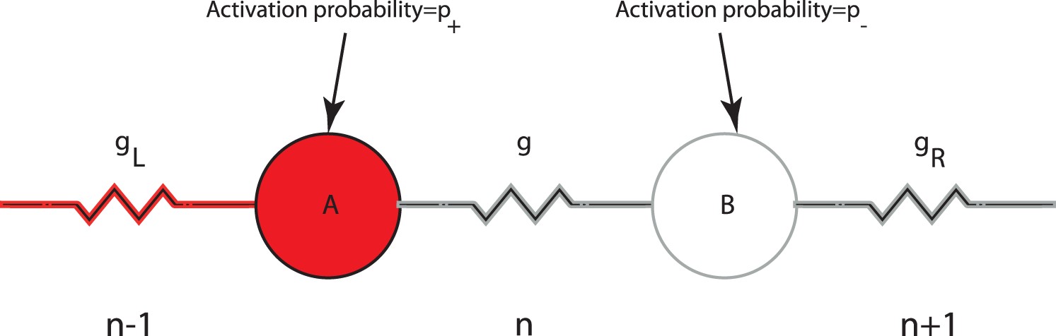
Figure 3. The bonds formalism of an islet.
-cells, A and B, are dominated by gap junctions
and
respectively. Each junction (
and
) can be in either strong (with probability
) or weak state (with probability
). A weak (strong) junction is likely to fire with a probability
(
). The central gap junction
is altered in response to the average potential difference of cells A and B,
, across it, according to a specified learning rule, such as the homeostatic rule of Fig. 2 that is considered here. For example, if cell A (red) here is assumed to fire in response to a strong
(this occurs with probability
) while cell B is silent (the probability with which it could have been active is
) in response to a weak
, then the bond, g, will be weakened since
. https://doi.org/10.1371/journal.pone.0070366.g003
There are few general principles that can organize an argument to discuss plastic behavior in excitable cells; Hebb’s postulate is one such. In common colloquialism this learning rule is stated as “cells that fire together, wire together”; in other words, temporal association between pairs of firing neurons is successively encoded in synaptic coupling between those neurons. A Hebbian philosophy asserts that the direction of adaptation is such as to reinforce coordinated activity between cells. One can now set forth some rules governing the above weight changes, which may have a Hebbian or anti-Hebbian flavor as the situation demands, and depend on the outcomes of the surrounding -cells. Hebbian rules in the case of synaptic plasticity favour synchrony, so that e.g. a synapse is strengthened if its surrounding neurons fire or do not fire together; the opposite is the case with anti-Hebbian rules. In the present context, we use this concept analogously: for Hebbian rules, synchronous activity causes a strengthening of conductance while anti-synchronous activity causes a weakening of conductance.Thus, loosely speaking, two gap junctions adjacent to any given gap junction “compete” to decide its type, and this continues to happen repeatedly across the entire network. Let us now consider the update dynamics of a single effective gap junction, that in some sense represents the average state of the whole network. To begin with, in such a picture, the outcomes are assumed to be uncorrelated at different locations, and treated as independent random variables, with the probability for activation being obtainable from the time-averaged activation frequency of the
-cell. Consistent with the situation described in the previous paragraph, that the effect of the common gap junction can be left out on average in comparing the outcomes of its connected
-cells, we associate, with each
-cell, a probability for activation at any instant that is only a function of the other neighboring gap junction, being equal to
(
) for a strong (weak) type gap junction.
We now consider a mean-field version of the model. The idea behind the mean-field approximation is that we look at the average behavior in an infinite system. This, at one stroke, deals with two problems: first, there are no fluctuations associated with system size, and second, the approximation that we have made in ignoring the “self-coupling” of the gap junction is better realized. In the mean-field representation, every gap junction is assigned a probability (uniform over the lattice) to be either strong () or weak (
), so that spatial variation is ignored, as are fluctuations and correlations. This single effective degree of freedom allows for a description of the system in terms of its fixed point dynamics. The rate of change of the probability
, say, (which in the limit of large system size is equivalent to the fraction of strong units) with time, is computed by taking into account only the nearest-neighbor gap junctional interactions, via specific rules.
To design a transition rule for gap junctions that is consistent with a Hebbian theory, and at the same time tunes gap junctional plasticity to voltage activity in the network, we mimic the homeostatic adaptation implicit in (fast) voltage-gating of conductance (Fig. 2): to reinforce synchronous activity conductance, changes must be directed towards a maximal state of conductance, while anti-synchronous activity is best served by a weakening of conductance. The homeostatic learning rule is summarised as follows: if -cells (Fig. 3) fire simultaneously
is zero and gap junction,
, strengthens to one, while if one
-cell fires but not the other,
is one and junction strength weakens to zero.
We write equations for the probability that the intermediate gap junction (Figure 3) is in the strong state, say, at time
in terms of the same probability at time
,
, the (complementary) probability that it was in the weak state at time
,
and Prob(
), the probability of a change in strength of a given magnitude.
The first term on the right hand side represents the probability that the strong state at time stays strong at time
; since the gap junctions are binary, a strong junction cannot get any stronger. Since
, this reduces to the equation
, independent of the initial state of the gap junction.
We now write down all possible scenarios for : in words, these correspond to the sum of the following probabilities: (Prob that both
and
are in the strong state)×(Prob that A and B both fire, AND both don’t fire)+(Prob that both
and
are in the weak state)×(Prob that A and B both fire, AND both don’t fire)+(Prob that
and
are in disparate states)×(Prob. that A and B both fire, AND both don’t fire).
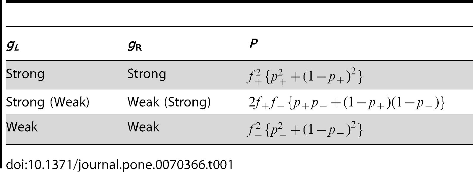
For example: if and
(see Fig. 3) are both strong – with probability
– the firing pattern that leads to a strong middle junction,
, according to the homeostatic learning rule is when
, i.e. either when both A and B fire simultaneously (probability,
), or both do not fire (probability,
). All such combinations are enumerated in Table 1, this leads to an equation for the evolution of
.
One major interest in developing a theory of gap junction adaption is to understand the changes in junctional conductance that take place in type 2 diabetes. It has been suspected from animal studies that loss of Cx36 is phenotypically similar to a prediabetic condition characterized by glucose intolerance, diminished insulin oscillations and first and second phases of insulin secretion, and a loss of beta-cell mass [6], [36]–[38]. Head et al. [11] have recently confirmed this in vivo via the observation that Cx36 conductance loss induces postprandial glucose intolerance in mice. These observations suggest that a loss of electrical connectivity in islets may underlie type 2 diabetes by disrupting insulin oscillations and reducing first-phase insulin secretion [6], [11].
If that electrical connectivity could be restored through the use of microcurrents, there is the very real possibility of not only treating type 2 diabetes but potentially reversing it.
Benninger et al. [39] have found yet another effect that could be relevant to diabetes, that a loss of gap junctions in islets leads to increased basal (i.e. when minimally stimulated by glucose) insulin release. If this were to hold in vivo it could explain hyperinsulinemia as a result of gap junction loss as well, when steady state levels of circulating plasma insulin in diabetics continue to be high even in fasting conditions.
Here again, the use of microcurrents that mimic our endogenous electrical connectivity could be used to treat hyperinsulinemia. Hyperinsulinemia is associated with hypertension, obesity, dyslipidemia, and glucose intolerance, a cluster of conditions collectively known as Metabolic syndrome. Using a microcurrent could potentially repair pancreatic beta-cell function thus helping those with type 1 diabetes impacted by hyperinsulinemia and aid those with type 2 diabetes whose body has become insulin resistant.
The game-theoretic formalism presented here provides a high-level explanation why a loss of junctional conductance would be necessary in diabetes. In the healthy individual insulin secretion occurs relatively sparingly, for a few hours at regularly spaced intervals following glucose ingestion (breakfast, lunch and dinner). The low firing rates in a healthy individual are accompanied by a high proportion of strong gap junctions (that is, near the region marked by A, Fig. 4, where is close to 1). Diabetes is associated with overnutrition among various other factors [40], and invariably involves combating an increased glucose load [41], [42]. Several authors that proposed that a substantial loss of Cx36 could occur in type 2 diabetes (reviewed for example in [37]). Much of the evidence that connexins expression or signaling are altered in models of type 2 diabetes comes from rodents; however, because Cx36 is present in human islets, this gives rise to the speculation (see e.g. [11]) that a loss of Cx36 gap junction conductance may occur in type 2 diabetes. Thus, based on glucose intolerance measured in the conscious mouse Head et al. [11], as well as others [5], [6], [43], [44], have estimated that a loss of nearly 50% in junctional conductance could occur in diabetes. In Fig. 4 the locus of a 50% connectivity loss is the line
, where the fraction of strong gap junctions is halved (
) but the firing rates are higher (Fig. 5). That is, the islet stressed by an increase glycemic stimulation is forced to respond with an increase in its firing and insulin secretion rate, which it does by degrading strong gap junctions to weaker ones.
In this way, the islet is able to accommodate a stimulus stronger than that for which its physiology had evolved. A change in is accomplished largely through altering the probabilities of junction-induced firing,
and
. As mentioned in the introduction, the classical view of diabetes is that it results from gap junction dysfunction. Instead, the game-theoretic theory we have presented relates a conductance decrease to an adaptive response of an islet that sacrifices strong gap junctions in order to maintain insulin control over hyperglycemia.
At the heart of our game-theoretic theory is its use of stochasticity in gap junction synchronisation. Classically, strong gap junctions entrain beta-cells to fire, the entire assembly is assumed to be fairly homogeneous in gap junction strength, and the resultant synchronous bursting is seen to be essential to GSIS. This theory on the other hand, introduces the possibility that beta-cells coupled even to strong gap junctions may not fire, and likewise, weak gap junctions may induce simultaneous firing. Further, synchronous bursting, as well as the simultaneous absence of bursting, induces stronger junctions, while anti synchrony weakens them. The result is that gap junctional strengths are constantly updated as a result of the synchronous or asynchronous bursting of beta-cells. In other words, the core idea of their paper is that disparate firing patterns lead to changes in gap junctional strength – which provides a hitherto unexplored scenario for synchrony. This then naturally leads to a situation where heterogeneity prevails in the distribution of gap junctional strengths in the islet. The heterogeneity of gap junctions in turn determines more complex patterns of activity in the network, beyond the simple categories of (anti-)synchronous bursting.
In principle it is possible to explain observations of junctional strengths such as in [39] individually, without recourse to a general theory of gap junction function. Typically, a lot of the focus is on studying the heterogeneity of beta-cells in an islet. Indeed, Benninger et al. verify that different thresholds exist for calcium excitations among the beta-cells of a (Cx36 null) islet, and conclude therefore that beta-cells with high thresholds create oscillator death [45] through gap junctions to decrease basal secretion. The other question to ask, however, is: can heterogeneous gap junctions within an islet shape the emergent properties of bursting? Once the heterogeneity of the gap junctions themselves is recognized as crucial, that leads, ipso facto, to an alternate view, one in which changes in junctional conductance are seen as solutions to an optimization problem. The essential ingredients of a theory of gap junction adaptation include keeping track of the propensities with which strong and weak junctions influence firing rates in beta-cells, and transition rules that determine how gap junctions will respond to local firing patterns. Here the central focus has been on learning the rules that embody homeostatic principles, which are a central feature of the energy maintenance pathways of the body.
The view that emerges instead is that the islet is sensitive to a glucose demand in secreting insulin and uses gap junctions as a tuning parameter in this adaptation. Thus looking to restore natural cellular function through the use of microcurrents or electrical connectivity repair holds much promise for the future of diabetes treatment, pancreatic islet cell repair and the potential reversal of type 2 diabetes.
References:
1.Goodenough DA, Paul DL (2009) Gap junctions. Cold Spring Harb Perspect Biol 1: a002576. View Article / Google Scholar
2.Nicholson BJ (2003) Gap junctions - from cell to molecule. J Cell Sci 116: 4479–4481. View Article / Google Scholar
3.Willecke K, Eiberger J, Degen J, Eckardt D, Romualdi A, et al. (2002) Structural and functional diversity of connexin genes in the mouse and human genome. Biol Chem 383: 725–737. View Article / Google Scholar
4.Winterhager E (2005) Springer-Verlag Berlin Heidelberg.
5.Ravier MA, Guldenagel M, Charollais A, Gjinovci A, Caille D, et al. (2005) Loss of connexin36 channels alters beta-cell coupling, islet synchronization of glucose-induced Ca2+ and insulin oscillations, and basal insulin release. Diabetes 54: 1798–1807. View Article / Google Scholar
6.Meda P (2012) The in vivo -to–cell chat room: connexin connections matter. Diabetes 61: 1656–1658. View Article / Google Scholar
7.Orci L, Unger RH, Renold AE (1973) Structural coupling between pancreatic islet cells. Experientia 29: 1015–1018. View Article / Google Scholar
8.Bosco D, Haefliger JA, Meda P (2011) Connexins: key mediators of endocrine function. Physiol Rev 91: 1393–1445. View Article / Google Scholar
9.Cabrera O, Berman DM, Kenyon NS, Ricordi C, Berggren PO, et al. (2006) The unique cytoarchitecture of human pancreatic islets has implications for islet cell function. Proc Natl Acad Sci USA 103: 2334–2339. View Article / Google Scholar
10.MacDonald PE, Rorsman P (2006) Oscillations, intercellular coupling, and insulin secretion in pancreatic beta cells. PLoS Biol 4: e49. View Article / Google Scholar
11. Head WS, Orseth ML, Nunemaker CS, Satin LS, Piston DW, et al. (2012) Connexin-36 gap junc-tions regulate in vivo first- and second-phase insulin secretion dynamics and glucose tolerance in the conscious mouse. Diabetes 61: 1700–1707. View Article / Google Scholar
12. Sherman A, Rinzel J, Keizer J (1988) Emergence of organized bursting in clusters of pancreatic beta-cells by channel sharing. Biophys J 54: 411–425. View Article / Google Scholar
13.Smolen P, Rinzel J, Sherman A (1993) Why pancreatic islets burst but single beta cells do not. The heterogeneity hypothesis. Biophys J 64: 1668–1680. View Article / Google Scholar
14.Sherman A, Smolen P (1997) Computer modeling of heterogeneous beta-cell populations. Adv Exp Med Biol 426: 275–284. View Article / Google Scholar
15.Bertram R, Sherman A, Satin LS (2010) Electrical bursting, calcium oscillations, and synchronization of pancreatic islets. Adv Exp Med Biol 654: 261–279. View Article / Google Scholar
16.Levin M, Stevenson CG (2012) Regulation of cell behavior and tissue patterning by bioelectrical signals: challenges and opportunities for biomedical engineering. Annu Rev Biomed Eng 14: 295–323. View Article / Google Scholar
17.Bertram R, Satin L, Zhang M, Smolen P, Sherman A (2004) Calcium and glycolysis mediate multiple bursting modes in pancreatic islets. Biophys J 87: 3074–3087. View Article / Google Scholar
18.Goel P, Sherman A (2009) The geometry of bursting in the dual oscillator model of pancreatic-cells. SIAM Journal on Applied Dynamical Systems 8: 1664–1693. View Article / Google Scholar
19.Meda P, Atwater I, Goncalves A, Bangham A, Orci L, et al. (1984) The topography of electrical synchrony among beta-cells in the mouse islet of Langerhans. Q J Exp Physiol 69: 719–735. View Article / Google Scholar
20.Bergsten P, Grapengiesser E, Gylfe E, Tengholm A, Hellman B (1994) Synchronous oscillations of cytoplasmic Ca2+ and insulin release in glucose-stimulated pancreatic islets. J Biol Chem 269: 8749–8753. View Article / Google Scholar
21.Lin JM, Fabregat ME, Gomis R, Bergsten P (2002) Pulsatile insulin release from islets isolated from three subjects with type 2 diabetes. Diabetes 51: 988–993. View Article / Google Scholar
22.Calabrese A, Zhang M, Serre-Beinier V, Caton D, Mas C, et al. (2003) Connexin 36 controls synchronization of Ca2+ oscillations and insulin secretion in MIN6 cells. Diabetes 52: 417–424. View Article / Google Scholar
23.Nunemaker CS, Zhang M, Wasserman DH, McGuinness OP, Powers AC, et al. (2005) Individual mice can be distinguished by the period of their islet calcium oscillations: is there an intrinsic islet period that is imprinted in vivo? Diabetes 54: 3517–3522. View Article / Google Scholar
24.Rocheleau JV, Walker GM, Head WS, McGuinness OP, Piston DW (2004) Microfluidic glucose stimulation reveals limited coordination of intracellular Ca2+ activity oscillations in pancreatic islets. Proc Natl Acad Sci USA 101: 12899–12903. View Article / Google Scholar
25.Stozer A, Gosak M, Dolensek J, Perc M, Marhl M, et al. (2013) Functional connectivity in islets of Langerhans from mouse pancreas tissue slices. PLoS Comput Biol 9: e1002923. View Article / Google Scholar
26.Camerer CF (2003) Behavioural studies of strategic thinking in games. Trends Cogn Sci 7: 225–231. View Article / Google Scholar
27.Szabo G, Fath G (2007) Evolutionary games on graphs. Physics Reports 446: 97–216. View Article / Google Scholar
28.Perc M, Szolnoki A (2010) Coevolutionary games A mini review. BioSystems 99: 109–125. View Article / Google Scholar
29.Mehta A, Luck JM (1999) Models of competitive learning: Complex dynamics, intermittent con-versions, and oscillatory coarsening. Phys Rev E 60: 5218–5230. View Article / Google Scholar
30.Mahajan G, Mehta A (2011) Competing synapses with two timescales as a basis for learning and forgetting. Europhys Lett 95: 109–125. View Article / Google Scholar
31.Bhat AA, Mahajan G, Mehta A (2011) Learning with a network of competing synapses. PLoS ONE 6: e25048. View Article / Google Scholar
32.Teubner B, Degen J, Sohl G, Guldenagel M, Bukauskas FF, et al. (2000) Functional expression of the murine connexin 36 gene coding for a neuron-specific gap junctional protein. J Membr Biol 176: 249–262. View Article / Google Scholar
33.Haefliger JA, Rohner-Jeanrenaud F, Caille D, Charollais A, Meda P, et al. (2013) Hyperglycemia downregulates Connexin36 in pancreatic islets via the upregulation of ICER-1/ICER-1. J Mol Endocrinol 51: 49–58. View Article / Google Scholar
34.Paulauskas N, Pranevicius H, Mockus J, Bukauskas FF (2012) Stochastic 16-state model of voltage gating of gap-junction channels enclosing fast and slow gates. Biophys J 102: 2471–2480. View Article / Google Scholar
35.Paulauskas N, Pranevicius M, Pranevicius H, Bukauskas FF (2009) A stochastic four-state model of contingent gating of gap junction channels containing two “fast” gates sensitive to transjunctional voltage. Biophys J 96: 3936–3948. View Article / Google Scholar
36.Bavamian S, Klee P, Britan A, Populaire C, Caille D, et al. (2007) Islet-cell-to-cell communication as basis for normal insulin secretion. Diabetes Obes Metab 9 Suppl 2118–132. View Article / Google Scholar
37.Hamelin R, Allagnat F, Haefliger JA, Meda P (2009) Connexins, diabetes and the metabolic syndrome. Curr Protein Pept Sci 10: 18–29. View Article / Google Scholar
38.Potolicchio I, Cigliola V, Velazquez-Garcia S, Klee P, Valjevac A, et al. (2012) Connexin-dependent signaling in neuro-hormonal systems. Biochim Biophys Acta 1818: 1919–1936. View Article / Google Scholar
39.Benninger RK, Head WS, Zhang M, Satin LS, Piston DW (2011) Gap junctions and other mechanisms of cell-cell communication regulate basal insulin secretion in the pancreatic islet. J Physiol (Lond) 589: 5453–5466. View Article / Google Scholar
40.Nathan DM, Buse JB, Davidson MB, Heine RJ, Holman RR, et al. (2006) Management of hyperglycemia in type 2 diabetes: A consensus algorithm for the initiation and adjustment of therapy: a consensus statement from the American Diabetes Association and the European Association for the Study of Diabetes. Diabetes Care 29: 1963–1972. View Article / Google Scholar
41.Inzucchi SE, Bergenstal RM, Buse JB, Diamant M, Ferrannini E, et al. (2012) Management of hyperglycaemia in type 2 diabetes: a patient-centered approach. Position statement of the American Diabetes Association (ADA) and the European Association for the Study of Diabetes (EASD). Diabetologia 55: 1577–1596. View Article / Google Scholar
42.Inzucchi SE, Bergenstal RM, Buse JB, Diamant M, Ferrannini E, et al. (2012) Management of hyperglycemia in type 2 diabetes: a patient-centered approach: position statement of the American Diabetes Association (ADA) and the European Association for the Study of Diabetes (EASD). Diabetes Care 35: 1364–1379.
43.Benninger RK, Zhang M, Head WS, Satin LS, Piston DW (2008) Gap junction coupling and calcium waves in the pancreatic islet. Biophys J 95: 5048–5061.
44.Speier S, Gjinovci A, Charollais A, Meda P, Rupnik M (2007) Cx36-mediated coupling reduces beta-cell heterogeneity, confines the stimulating glucose concentration range, and affects insulin release kinetics. Diabetes 56: 1078–1086. View Article / Google Scholar
45.Bar-Eli K, Ermentrout B (2008) Oscillation death. Scholarpedia 3: 5371.
46. Goel P, Mehta A (2013) Learning Theories Reveal Loss of Pancreatic Electrical Connectivity in Diabetes as an Adaptive Response. PLoS ONE 8(8): e70366.


Comments