CHECK SOURCE OPERATING PROCEDURE
Prior to performing a radiation survey using the survey meter, the RHSO must first ensure that the instrument is functioning properly. The RHSO must perform an operational check using the check source (Strontium-90) that is affixed to the side of the instrument. The location of the check source is marked with "Caution - Radioactive Material" (CRAM) tape and an "X" to indicate where to position the probe when performing the operational check.
One of the emerging techniques in prostate cancer imaging is magnetic resonance spectroscopy (MRS), which provides bio-chemical information from body tissue in a safe and noninvasive method. In the presence of an applied external magnetic field, the atomic nuclei within molecules behave like tiny magnets and resonate at different frequencies at the same magnetic field strength. This is the basic principle of magnetic resonance imaging (MRI) and spectroscopy (1-4).
Proton Therapy (PT) is a high-quality radiation therapy and initially used to treat human patients in Lawrence Berkeley Laboratory. For about 50 years of its use in treating cancer, it has been reported to have an excellent clinical result especially in prostate cancer (Smith, 2006). The precise dose deposition to the tumor volume prescribed in a cancer patient (Kim, 2006) improved local control and a disease-free survival (Smith, 2006).
What other scientists say
Sample Radiation Protection and Safety/ALARA Program based on Philippine Perspective
Statement from the management.
I. Organization and ResponsibilitiesChair
Vice-Chair
Board members
Personnel
Authorized user
Radiation protection officer
Supervisor
Medical physicist
Radiation therapist
Moulding technologist
Radiation oncology nurse
Positron Emission Tomography (PET) was invented at the Mallinckrodt Institute of Radiology at Washington University in the mid 1970s and was used as a research tool in neurology, cardiology, and oncology (1).
It has been developed as a tool for imaging and quantification of cellular and molecular processes in vivo and made an important role in staging diseases and monitoring response to treatment (2).
In comparison to anatomic imaging modalities, PET produces images of biochemical and physiologic processes in tissues that help distinguishing benign and malignant lesions when CT and MRI cannot (1,3) .

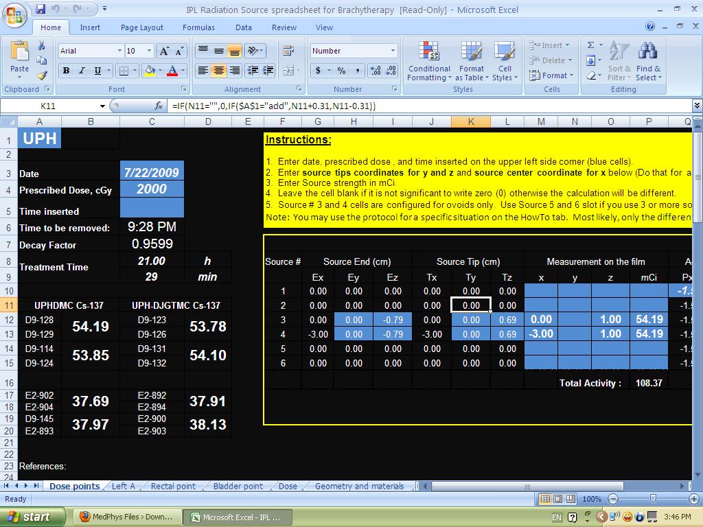 LDR Brachytherapy Verification Spreadsheet
 Prostate Cancer Imaging and Radiation Treatment Modalities
 Lesson from the past: Radiation Therapy Overexposure